- Sponsored Content
Ask the Expert - 2D DIGE Western Blots for HCP Detection and Coverage Determination: Advantages of Fluorescence over Colorimetric Detection
March 10, 2015
Sponsored by BioGenes
with Jamina Fiedler, BioGenes GmbH
Biopharmaceutical products must be free of by-products (such as host-cell proteins, HCPs) for approval by authorities such as the US FDA. For that, a clear proof is required of sufficient HCP detection by the HCP assay used. The two-dimensional (2D) DIGE Western blot method is the standard technique for detection of HCP coverage. Colorimetric detection has several limitations that can be overcome by the use of fluorescent detection.
Jamina’s Presentation
Practical differences between 2D DIGE Western blotting and a colorimetric procedure are the DIGE labeling of samples before their application and the use of fluorescent-labeled antibodies. All other steps such as sample preparation, isoelectric focusing (IEF), sodium dodecyl sulfate (SDS) polyacrylamide gel electrophoresis (PAGE), and Western blot transfer are the same. We work with GE Healthcare equipment and 20 × 26 cm (~7.9 × 10 in) gels. That large gel size improves resolution.
The first advantage of fluorescence is the high sensitivity, which is very important for small sample amounts. Comparing a Coomassie gel of 600 µg with a CyDye blot of 50 µg shows similar intensity. But the CyDye blot contains <10% the amount of sample.
The second advantage is the fluorescent method’s high dynamic range. The high sensitivity and broad dynamic range of the CyDye DIGE fluors enable detection of both abundant and scarce proteins. The evaluation software uses the whole summation of the total dynamic range. The high dynamic range applies to the patterns of HCP and antibody detection. Therefore, you do not need to produce multiple blots with different intensities.
The third advantage is overcoming intergel variation. In the usual colorimetric procedure, two gels are run. One gel is used for total protein staining by Coomassie or silver, and the other is for Western blotting with immunostain. Variation between the two gels makes spot matching necessary for evaluation. That can be difficult, and inexperienced users can make mistakes.
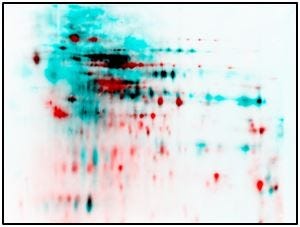
The first step in the new fluorescence procedure is labeling a sample with CyDye DIGE Fluor minimal dye. Then you run only one gel with subsequent Western blotting and immunostaining using fluorescent antibodies. That results in a 2D DIGE Western blot membrane on which the HCP and antibodies can be detected.
One result of eliminating intergel variation is that no matching is necessary. Also, transfer efficiency has no influence on coverage determination. And 2D DIGE Western blotting facilitates HCP detection and makes complete electronic evaluation possible. Intergel distortions are no problem.
Questions and Answers
What is the pH range of the IEF strips?
We use a pH range of 3–10 to show a complete overview of the full pH range on a 2D gel.
What software do you use to determine HCP coverage?
We use ImageMaster from GE Healthcare.
Does each exposure during image capture reduce the intensity of fluorescence? Does it get lighter over multiple times without fluorescence?
No, I can scan the membrane again and again. I haven’t determined whether it gets less bright over 20 times. I’ve never done it so often to get less fluorescence over time. I store them in the dark, so I can scan them again months later.
Do you find any difference between wet transfer and semidry membrane transfer? It’s difficult to do wet transfer on such a big gel. So we haven’t done tank blotting with these gels. We’ve always done semidry blotting.
How do you calculate the sequence coverage of an antibody if you have areas of high saturation?
That can occur with colorimetric staining and makes multiple blots necessary for proper evaluation. To obtain at least two different intensities you would choose, for example, one blot with a small antigen and antibody amount and a second blot with a large amount of antigen and antibody. In fluorescence, you don’t have this problem because the dynamic range is high enough to prevent overstaining blur.
Does the fluorescent label change the 2D pattern?
It does not. The CyDye DIGE minimal labeling is designed by GE Healthcare, and we have not seen any shift in the isoelectric point of a protein. Because the label is so small, it has no effect on protein size.
More Online
The full presentation of this “Ask the Expert” webcast — with data — can be found on the BioProcess International website at http://bit.ly/BioGenes-ATE
You May Also Like


